🔍 1. “A Cyst Giving the Evil Eye”? — What Does This Mean?
This is not a formal medical term, but rather a vivid, colloquial (and often humorous) description used by dermatologists, estheticians, and even patients to describe a very specific clinical appearance:
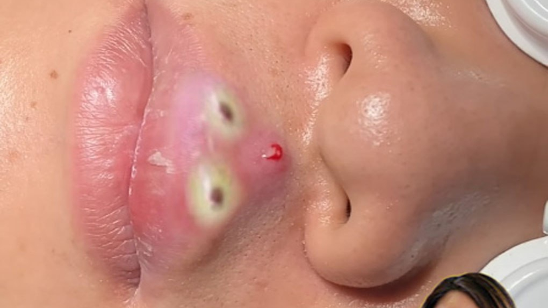
✅ A large, tense, dome-shaped epidermal (infundibular) cyst — typically on the eyelid, brow, or temple — that has:
- A central punctum (tiny dark opening, like a “pupil”),
- A smooth, glistening, skin-colored or yellowish dome (the “sclera”),
- And is positioned so it seems to stare directly at you — especially when the patient looks straight ahead.
👁️ Why “Evil Eye”?
- The cyst’s round, bulging shape + dark central punctum resembles a cartoonish or eerie eye.
- When located near the medial canthus (inner eye corner), it can appear to “follow” you as the patient moves — enhancing the uncanny effect.
📌 Real-World Example:
A 45-year-old patient presents with a 1.2 cm firm, mobile nodule on the upper eyelid, with a central black dot. On examination, it’s a classic epidermal inclusion cyst — but everyone in the clinic jokes: “It’s giving the evil eye!”
📚 2. More Detail with Reference
✅ Medical Identity:
This is almost always an epidermal (infundibular) cyst (formerly called sebaceous cyst — a misnomer, as it’s not sebaceous in origin).
📌 Key Reference:
Bolognia JL, Schaffer JV, Cerroni L (eds.). Dermatology, 5th ed. Elsevier, 2023.
— Chapter 82: Cysts and Pseudocysts of the Skin
— Confirms: >95% of “sebaceous cysts” are actually epidermal inclusion cysts; true sebaceous cysts (steatocystoma) lack a granular layer and contain oil, not keratin.
📌 Supporting Evidence:
Zemtsov A, et al. Eyelid cysts: Clinical and histopathologic correlation. Dermatol Surg. 1998;24(11):1165–1168.
— Found epidermal cysts accounted for 62% of excised eyelid cysts; all had central puncta and “domed” morphology.
💊 3. Solutions & Treatments
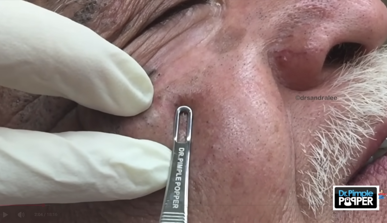
⚠️ Do NOT attempt extraction at home — periorbital skin is thin, highly vascular, and close to the eye. Risk of infection, scarring, or orbital cellulitis is significant.
✅ Definitive Treatment: Complete Surgical Excision
📌 Success Rate: >95% cure with intact excision (Zemtsov A, 1998).
📌 Recurrence: Usually due to rupture during extraction → granulomatous inflammation + incomplete removal.
🌿 Non-Surgical / Conservative Options (Temporary or Palliative)
🚫 Avoid:
- Incision & drainage (I&D) alone — high recurrence
- “Expressing” the cyst — almost always ruptures the wall
- Topical retinoids/antibiotics — do not resolve established cysts