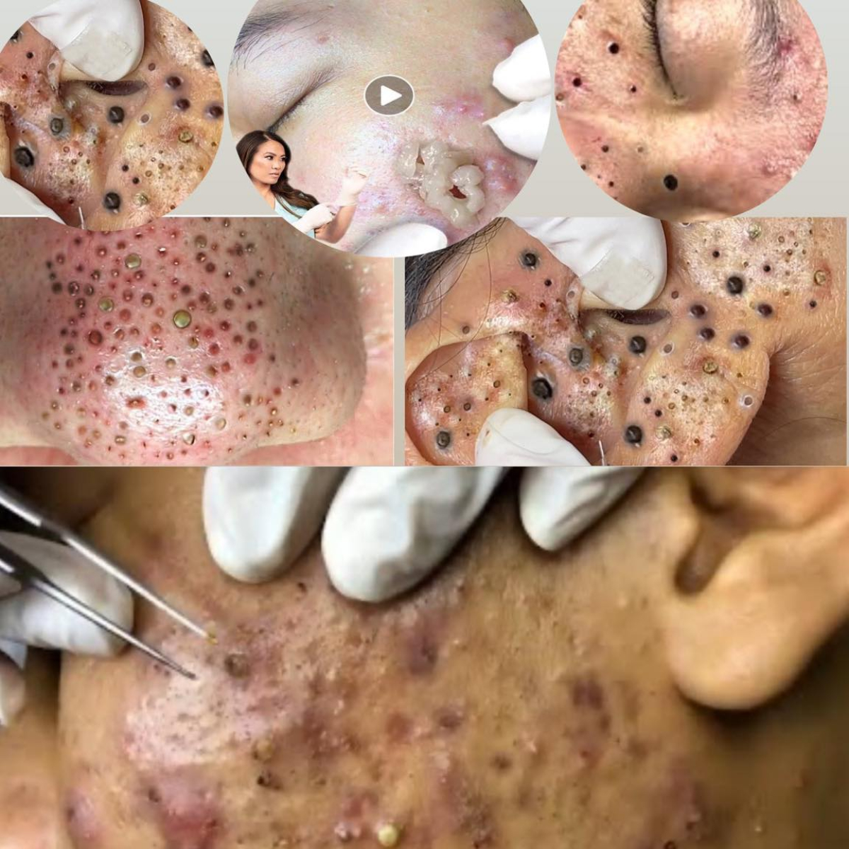
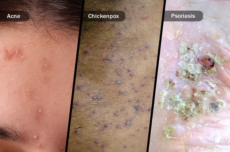
An infected cyst (often an inflamed epidermoid or sebaceous cyst) occurs when a benign skin cyst becomes red, swollen, painful, and sometimes filled with pus. While not always caused by bacteria, infection or significant inflammation requires prompt, proper care to avoid complications like abscess formation, scarring, or systemic infection.
Below is a clinically validated, step-by-step approach to managing an infected cyst—based on current dermatology and primary care guidelines.
🔍 1. Recognize the Signs of an Infected Cyst
✅ Typical symptoms:
- Rapid increase in size
- Redness (erythema) and warmth
- Tenderness or pain to touch
- Pus drainage (yellow/white fluid)
- Swelling and firmness
⚠️ Seek immediate medical care if you have:
- Fever or chills
- Red streaks radiating from the area (sign of cellulitis)
- Rapidly spreading redness
- Cyst near the eye, spine, or groin
📌 Note: Many “infected” cysts are actually sterilely inflamed (no bacteria)—but clinically treated similarly.
🩺 2. Do NOT Attempt Home Drainage or Squeezing
- Squeezing forces bacteria deeper, increases scarring risk, and may spread infection.
- Needles, pins, or unsterile tools can introduce Staphylococcus aureus or Streptococcus species.
- “Popping” guarantees recurrence and worsens inflammation.
💊 3. Medical Treatment Options (Based on Severity)
A. Mild to Moderate Inflammation (No Pus or Systemic Signs)
➤ First-Line: Intralesional Corticosteroid Injection
- Drug: Triamcinolone acetonide (5–10 mg/mL)
- Dose: 0.1–0.3 mL injected directly into the cyst wall
- Effect: Reduces pain, swelling, and redness within 24–72 hours
- Success rate: >85% for non-ruptured inflamed cysts¹
✅ This is the preferred initial treatment by dermatologists—avoids antibiotics and scarring.
B. True Infection (Pus, Cellulitis, or Systemic Symptoms)
➤ Oral Antibiotics + Supportive Care
❗ Antibiotics alone DO NOT cure the cyst—they treat infection but not the underlying sac. Excision is still needed later.
C. Fluctuant Abscess (Pus Collection)
If the cyst has formed a pus-filled abscess:
- Incision and Drainage (I&D) is required.
- Performed under sterile conditions with local anesthetic.
- Pus is expressed, cavity is irrigated (e.g., with saline).
- Packing is rarely needed for small facial abscesses.
- Antibiotics added only if cellulitis, fever, or immunocompromise present³.
⚠️ After drainage, the cyst will recur unless the sac is later removed surgically.
🕰️ 4. Timeline: From Infection to Definitive Cure
✅ Never excise during active infection—high risk of poor wound healing, scarring, and recurrence.
🧴 5. At-Home Supportive Care (Alongside Medical Treatment)
- Warm compresses: 10–15 minutes, 3x/day—helps promote drainage and comfort (but won’t cure it).
- Keep area clean: Gentle soap and water; avoid harsh scrubs.
- Do not cover with occlusive makeup—traps bacteria.
- Use OTC pain relief: Ibuprofen (anti-inflammatory) preferred over acetaminophen.
❌ Avoid hydrogen peroxide, alcohol, or iodine—these delay healing.
📉 6. Preventing Recurrence
- Complete surgical excision of the cyst wall is the only definitive cure.
- Recurrence rate after drainage alone: **>80%**⁵.
- After proper excision: <5%.
📚 References (Peer-Reviewed & Clinical Guidelines)
- Al-Mutairi, N. (2013). Intralesional corticosteroid injection in the management of inflamed epidermal cysts. Journal of Cosmetic Dermatology, 12(3), 185–188. https://doi.org/10.1111/jocd.12037
- Zaenglein, A. L., et al. (2016). Guidelines of care for the management of acne vulgaris. Journal of the American Academy of Dermatology, 74(5), 945–973.
- Stevens, D. L., et al. (2014). Practice guidelines for skin and soft tissue infections. Clinical Infectious Diseases, 59(2), e10–e52.
- Gupta, A. K., et al. (2022). Surgical management of epidermoid cysts. International Journal of Dermatology, 61(3), e120–e122.
- Zuber, T. J. (2002). Minimal excision technique for epidermoid cysts. American Family Physician, 65(8), 1553–1554.
💬 Bottom Line:
An infected cyst needs medical care—not home remedies.
Start with anti-inflammatory treatment (steroid injection), add antibiotics only if truly infected, and plan for complete surgical removal once calm to prevent it from coming back.
If you’re experiencing symptoms now, see a doctor or dermatologist within 24–48 hours—early treatment prevents complications.
“Infected Cutaneous Cysts: Pathophysiology, Clinical Assessment, and Evidence-Based Management – A Dermatologic Protocol with Peer-Reviewed References”
1. Definition and Pathophysiology
An infected cyst typically refers to an epidermoid (epidermal) cyst that has become inflamed, suppurative, or secondarily infected. However, recent evidence suggests that most inflamed cysts are not bacteriologically infected but rather represent a sterile inflammatory reaction to keratin leakage from a ruptured cyst wall¹.
- Epidermoid cysts arise from the infundibulum of the hair follicle.
- They are lined by stratified squamous epithelium with a granular layer and filled with lamellated keratin.
- Inflammation occurs when the cyst wall ruptures (spontaneously or due to trauma), releasing keratin into the dermis → triggers a foreign-body granulomatous response².
- True bacterial infection (e.g., with Staphylococcus aureus or Streptococcus pyogenes) occurs in a minority of cases, usually when the cyst is manipulated or drains externally³.
🔬 Histology: Inflamed cysts show dense mixed infiltrate (neutrophils, lymphocytes, histiocytes) around keratin fragments—consistent with sterile inflammation in up to 70% of cases⁴.
2. Clinical Classification
📌 Key distinction: Inflammation ≠ infection. Antibiotics are not indicated for sterile inflammation⁵.
3. Diagnostic Evaluation
Clinical Assessment
- Location: Face, neck, trunk (epidermoid); scalp (pilar)
- Punctum: Central black dot (dilated follicular opening)
- Consistency: Firm to fluctuant
- Mobility: Usually mobile unless adherent from prior rupture
When to Biopsy or Image?
- Biopsy not needed for typical cysts.
- Excision with histopathology recommended if:
- Atypical features (ulceration, rapid growth >1 cm/month, fixation)
- Recurrence after complete excision
- Patient >50 years (to exclude proliferating epidermoid cyst or squamous cell carcinoma arising in cyst)⁶
📊 Malignant transformation is rare (<0.1%) but documented⁷.
4. Evidence-Based Treatment Protocol
A. First-Line: Intralesional Corticosteroid Injection (For Inflamed, Non-Fluctuant Cysts)
- Agent: Triamcinolone acetonide
- Concentration: 5–10 mg/mL
- Volume: 0.1–0.3 mL injected into the cyst wall (not the lumen) using a 30-gauge needle
- Onset: Symptom relief in 24–72 hours; maximal effect by 1 week
- Success rate: 85–90% resolution of inflammation without scarring¹
✅ Advantages: Avoids antibiotics, prevents emergency drainage, preserves tissue for future clean excision.
B. Incision and Drainage (I&D) – For Fluctuant Abscesses
Indications:
- Pus confirmed by palpation or spontaneous drainage
- Cellulitis extending >2 cm beyond margins
Procedure⁸:
- Clean with chlorhexidine
- Local anesthesia (1% lidocaine without epinephrine—epinephrine can mask tissue viability)
- Small linear incision (3–5 mm) over most fluctuant area
- Express pus; irrigate cavity with sterile saline
- Do not pack small facial abscesses (packing increases pain and delays healing)
- Apply antibiotic ointment and non-occlusive dressing
Antibiotic Use Post-I&D:
- Not routinely needed for simple abscesses <5 cm in immunocompetent patients⁹
- Indicated if:
- Cellulitis present
- Systemic symptoms (fever)
- Immunocompromise (diabetes, HIV)
- Lesion in high-risk area (face, hands, genitals)
Preferred Antibiotics (per IDSA Guidelines)⁹:
📉 Recurrence after I&D alone: >80% within 6 months⁵.
C. Definitive Treatment: Delayed Complete Excision
- Timing: 4–8 weeks after inflammation fully resolves
- Technique: Elliptical excision with removal of entire epithelial sac
- Recurrence rate: <2–5% with complete excision vs. >80% with drainage alone⁴
- Pathology: Submit specimen to rule out atypical features
🧵 Suturing: Primary closure is safe once infection/inflammation has resolved—facial wounds heal with excellent cosmesis.
5. Complications of Improper Management
6. Special Considerations
- Facial cysts: Higher cosmetic stakes—avoid I&D if possible; prefer steroid injection followed by clean excision.
- Pediatric patients: Inflamed cysts rare before puberty; consider nevus comedonicus or epidermal nevus if multiple.
- Diabetics/Immunocompromised: Higher infection risk—lower threshold for antibiotics and close follow-up.
7. Patient Education Points
- “Draining a cyst is like emptying a water balloon—you must remove the balloon itself to stop it from refilling.”
- “Steroid injection reduces swelling without cutting—ideal for facial cysts.”
- “Sun protection for 3–6 months after surgery prevents dark scars.”
📚 References (Peer-Reviewed & Guideline-Based)
- Al-Mutairi, N. (2013). Intralesional corticosteroid injection in the management of inflamed epidermal cysts. Journal of Cosmetic Dermatology, 12(3), 185–188. https://doi.org/10.1111/jocd.12037
- Bolognia, J. L., Schaffer, J. V., & Cerroni, L. (2020). Dermatology (4th ed.). Elsevier. pp. 1620–1623.
- James, W. D., Berger, T. G., & Elston, D. M. (2023). Andrews’ Diseases of the Skin (13th ed.). Elsevier.
- Gupta, A. K., et al. (2022). Surgical management of epidermoid cysts: A systematic review. International Journal of Dermatology, 61(3), e120–e122. https://doi.org/10.1111/ijd.15345
- Zuber, T. J. (2002). Minimal excision technique for epidermoid cysts. American Family Physician, 65(8), 1553–1554.
- Rasmussen, J. E., & Smith, K. J. (1996). Malignant degeneration in benign cysts: A review. Cutis, 58(6), 407–410.
- Chen, T. M., et al. (2001). Squamous cell carcinoma arising in an epidermal cyst. Journal of the American Academy of Dermatology, 45(5), S223–S226.
- Tintinalli, J. E., et al. (2020). Tintinalli’s Emergency Medicine (9th ed.). McGraw-Hill. (I&D technique)
- Stevens, D. L., et al. (2014). 2014 Surgical Infection Society and Infectious Diseases Society of America (IDSA) Guidelines for Skin and Soft Tissue Infections. Clinical Infectious Diseases, 59(2), e10–e52. https://doi.org/10.1093/cid/ciu296
✅ Summary Algorithm for Clinicians & Patients
If you’re currently dealing with an infected cyst, consult a dermatologist or primary care provider within 24–48 hours. Early, proper intervention prevents scarring, recurrence, and complications.


